No products in the cart.
Internal Medicine, Neurology, Urology
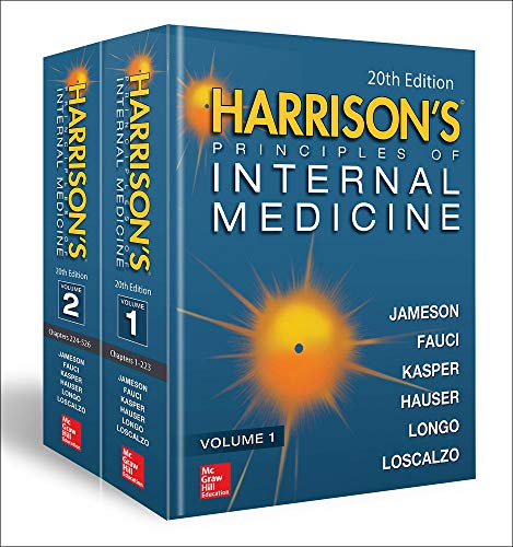
Harrison’s Principles of Internal Medicine, 20th Edition (Complete Videos Set)
180 Videos
File Size = 8.40 GB
$20.00
[
Video list:
Video 027-1: Classic Cataplexy
Video 027-2: REM Sleep Behavior Disorder
VIDEO 236-1: Cine steady-state free precession (SSFP) imaging (left) in short axis in a patient who had a large anterior myocardial infarction.
VIDEO 236-2: Cine cardiac magnetic resonance (CMR) imaging of a patient in the long-axis four-chamber view.
VIDEO 236-3: Exercise echocardiogram showing rest images on left and poststress images on right, with parasternal long-axis, upper panel, and apical four-chamber, lower panel, end-systolic frames.
VIDEO 236-4: Cardiac magnetic resonance (CMR) myocardial perfusion imaging during vasodilating stress, in three parallel-short-axis views.
VIDEO 236-5: A 60-year-old female presented with intermittent chest pain of 3 days in duration but was pain free at the time of assessment in the emergency room.
VIDEO 236-6: A patient with severe aortic regurgitation quantified by cardiac magnetic resonance (CMR).
VIDEO 236-7: T2* images of the heart (left panel) and the liver (right panel) of a patient who has hemochromatosis.
VIDEO 236-8: The heart in long and short axis.
VIDEO 285-1: Cystic Fibrosis
VIDEO 352-1A: Transthoracic echocardiographic images of a 9-year-old girl with first episode of acute rheumatic fever.
VIDEO 352-1B: Transthoracic echocardiographic images of a 9-year-old girl with first episode of acute rheumatic fever.
VIDEO 352-1C: Transthoracic echocardiographic images of a 9-year-old girl with first episode of acute rheumatic fever.
VIDEO 352-1D: Transthoracic echocardiographic images of a 9-year-old girl with first episode of acute rheumatic fever.
VIDEO 352-2A: Transthoracic echocardiographic images are from a 5-year-old boy with chronic rheumatic heart disease with severe mitral valve regurgitation and moderate mitral valve stenosis.
VIDEO 352-2B: Transthoracic echocardiographic images are from a 5-year-old boy with chronic rheumatic heart disease with severe mitral valve regurgitation and moderate mitral valve stenosis.
VIDEO 440-1: Myasthenia Gravis and Other Diseases of the Neuromuscular Junction
VIDEO A08-01: Echocardiography in the evaluation of acute myocardial infarction (parasternal long-axis view).
VIDEO A08-02: Echocardiography in the evaluation of acute myocardial infarction (apical four-chamber view).
VIDEO A08-03: Stress echocardiography for suspected CAD (rest apical four-chamber view).
VIDEO A08-04: Stress echocardiography for suspected CAD (stress apical four-chamber view).
VIDEO A08-05: CMR stress myocardial perfusion images (short-axis cine images).
VIDEO A08-06: CMR stress myocardial perfusion images (short-axis perfusion images).
VIDEO A08-07: ECG gated images.
VIDEO A08-08: A case of viability assessment in a patient with inferior myocardial infarction (short-axis cine images).
VIDEO A08-09: Echocardiography for evaluation of the severity of aortic stenosis (parasternal long-axis view).
VIDEO A08-10: Echocardiography for evaluation of the severity of aortic stenosis (parasternal long-axis view).
VIDEO A08-11: Echocardiography for evaluation of the severity of aortic stenosis (short-axis view).
VIDEO A08-12: Utility of echocardiography to evaluate for cardiac amyloidosis (parasternal long axis view).
VIDEO A08-13: Utility of echocardiography to evaluate for cardiac amyloidosis (short-axis view).
VIDEO A08-14: Utility of echocardiography to evaluate suspected hypertrophic cardiomyopathy (parasternal long-axis view).
VIDEO A08-15: Utility of echocardiography to evaluate suspected hypertrophic cardiomyopathy (apical four-chamber view).
VIDEO A08-16: Utility of echocardiography to evaluate suspected hypertrophic cardiomyopathy (Doppler apical four-chamber view).
VIDEO A08-17: Classic MR findings in a patient with hypertrophic cardiomyopathy (short-axis cine images).
VIDEO A08-18: Utility of echocardiography to evaluate cardiac involvement in cancer (parasternal long-axis view).
VIDEO A08-19: Classic echocardiographic findings in pulmonary hypertension (parasternal long-axis view).
VIDEO A08-20: Classic echocardiographic findings in pulmonary hypertension (short-axis view).
VIDEO A08-21: Metastatic cardiac tumor (four-chamber view with MRI).
VIDEO A08-22: Metastatic cardiac tumor (PET whole-body cine view).
VIDEO A10-1: Pulse Pressure.
VIDEO A10-2: Plaque Instability and Acute Events.
VIDEO A10-3: The Lipoprotein Menagerie.
VIDEO A10-4: Pathogenesis of the Atheroscleratic Plaque and Acute Coronary Syndromes
VIDEO A10-5: Atherogenesis.
VIDEO A10-6: Metabolic Syndrome, Diabetes and Atherogenesis.
VIDEO A11-01: Baseline left coronary angiogram shows an occluded LCx with left-to-left collaterals originating from LAD septal vessels.
VIDEO A11-02: Attempts to cross the total occlusion in the LCx using a hydrophilic wire and an antegrade approach were not successful with the wire tracking to the right of the trajectory.
VIDEO A11-03: The LAD septal collateral is accessed with a guidewire that is directed toward the distal LCx to cross the total occlusion retrograde.
VIDEO A11-04: The total occlusion is crossed retrograde. The wire is snared in the guide, exteriorized, and used to provide antegrade access to the LCx.
VIDEO A11-05: Antegrade flow in the LCx is restored after balloon inflation.
VIDEO A11-06: Following stenting of the total occlusion, blood flow in the distal vessel is improved and a second significant stenosis is seen.
VIDEO A11-07: Final result after LCx stenting.
VIDEO A11-08: Baseline angiogram of the left coronary circulation shows the significant stenosis in the mid-LAD and the bifurcation lesion involving a large diagonal branch.
VIDEO A11-09: Both vessels are accessed with guidewires and pretreated with balloon angioplasty.
VIDEO A11-10: Result after balloon angioplasty.
VIDEO A11-11: Stent being positioned in the LAD.
VIDEO A11-12: LAD poststent result.
VIDEO A11-13: Stent deployed in the diagonal branch through the stent struts in the LAD using the “culotte” technique.
VIDEO A11-14: Diagonal branch poststent result.
VIDEO A11-15: Simultaneous inflation of two 2.5-mm “kissing” balloons.
VIDEO A11-16: Final postbifurcation stenting result.
VIDEO A11-17: The right coronary artery (RCA) is totally occluded with filling defects in the vessel after contrast injection, indicating thrombus is present in the vessel.
VIDEO A11-18: An angioplasty wire is threaded through the thrombotic lesion, but this does not restore blood flow to the distal vessel.
VIDEO A11-19: Result after manual thrombectomy and thrombus extraction.
VIDEO A11-20: After balloon angioplasty and stenting, thrombus is still present.
VIDEO A11-21: After repeat manual thrombectomy and expansion of the stent, the thrombus is no longer present.
VIDEO A11-22: Final result.
VIDEO A11-23: Saphenous vein graft to a first obtuse marginal branch with an 80% eccentric stenosis in the midgraft.
VIDEO A11-24: A distal protection device is deployed past the lesion.
VIDEO A11-25: Angioplasty balloon inflation with the distal protection device in place.
VIDEO A11-26: Final result after stent placement.
VIDEO A11-27: Baseline left coronary artery injection in right anterior oblique (RAO) cranial projection shows a high-grade calcified stenosis in the left main coronary artery and a significant stenosis in the proximal LAD.
VIDEO A11-28: In the left anterior oblique (LAO) caudal view, the left main coronary artery lesion can be seen to extend into the ostia of both the LCx and the LAD.
VIDEO A11-29: Guidewires were placed into both the LCx and LAD.
VIDEO A11-30: The bifurcation lesion in the left main coronary artery extending into the LCx and LAD ostia is treated using a “culotte” technique.
VIDEO A11-31: Next, the LAD wire is removed and passed through the stent into the distal LAD.
VIDEO A11-32: Following rewiring of the LCx, both stents are re-dilated simultaneously (“kissing” balloons).
VIDEO A11-33: The final result in the LAO caudal view.
VIDEO A11-34: The final result in the RAO cranial view showing patent left main, LCx, and LAD coronary arteries.
VIDEO A11-35: Baseline angiogram of the left coronary circulation in the RAO view shows the total occlusion of the second obtuse marginal branch with delayed retrograde filling via collateral vessels and a high-grade stenosis in the ramus intermedius.
VIDEO A11-36: A guidewire is passed through the total occlusion, and the lesion is pretreated with balloon angioplasty.
VIDEO A11-37: Following placement of a drug-eluting stent in the lesion, the vessel is widely patent.
VIDEO A11-38: The ramus intermedius lesion was crossed with a guidewire and pretreated with balloon angioplasty.
VIDEO A11-39: A drug-eluting stent is placed across the ramus lesion and deployed.
VIDEO A11-40: Baseline angiogram of the RCA shows a high-grade lesion in the midsegment of the vessel.
VIDEO A11-41: The lesion was pretreated with balloon dilation followed by stent deployment.
VIDEO A11-42: The final result shows no residual stenosis in the mid-RCA.
VIDEO A11-43: Baseline angiogram of the RCA in an anterior-posterior (AP) cranial view showing a tortuous vessel with a stenosis in the proximal vessel.
VIDEO A11-44: Baseline angiogram of the left coronary circulation showing in-stent restenosis in the proximal LCx stent.
VIDEO A11-45: A pressure wire is advanced past the in-stent restenosis lesion in the LCx, intravenous adenosine if given to achieve maximal hyperemia, and the fractional flow reserve (FFR) is measured.
VIDEO A11-46: After a washout period, adenosine is infused again and FFR is measured in the RCA.
VIDEO A11-47: Following placement of drug-eluting stents in the RCA, there is no residual stenosis in the vessel.
VIDEO A11-48: Baseline angiogram showing a total occlusion of the proximal LAD within the drug-eluting stent and a significant stenosis at the origin of the LCx.
VIDEO A11-49: The LAO view shows the LCx stenosis with a filling defect indicating that thrombus is present in the vessel lumen.
VIDEO A11-50: The LAD lesion was crossed with a guidewire, which resulted in slow filling of the mid-LAD (TIMI 2 flow) and revealed thrombus filling the stent.
VIDEO A11-51: The final result after LAD and LCx stenting.
VIDEO A11-52: Aortogram shows patent coronary arteries and minimal aortic insufficiency.
VIDEO A11-53: Balloon valvuloplasty is performed with rapid ventricular pacing at 180 beats/min.
VIDEO A11-54: A 26-mm Edwards SAPIEN valve is positioned using fluoroscopic and transesophageal echo guidance and deployed.
VIDEO A11-55: Aortogram after valve deployment shows a functional valve with mild aortic insufficiency and without impingement of the coronary ostia.
VIDEO A11-56: A sizing balloon is placed across the ASD.
VIDEO A11-57: An Amplatzer® septal occluder is being positioned across the ASD.
VIDEO A11-58: The two disks of the device in place across the ASD.
VIDEO A12-1: Normal Ultrasound.
VIDEO A12-2: ARDS (Ultrasound).
VIDEO A12-3: Pneumothorax.
VIDEO A12-4: Pleural Effusion.
VIDEO CP01-1: Clinical Procedure Tutorial: Central Venous Catheter Placement.
VIDEO CP02-1: Clinical Procedure Tutorial: Thoracentesis.
VIDEO CP03-1: Clinical Procedure Tutorial: Abdominal Paracentesis.
VIDEO CP04-1: Clinical Procedure Tutorial: Endotracheal Intubation.
VIDEO CP05-1: Clinical Procedure Tutorial: Percutaneous Arterial Blood Gas Sampling.
VIDEO CP06-1: Clinical Procedure Tutorial: Lumbar Puncture.
VIDEO CP07-1: Clinical Procedures Tutorial: Phlebotomy.
VIDEO CP08-1: Clinical Procedures Tutorial: Insertion of Female Urethral Catheter.
VIDEO CP09-1: Clinical Procedures Tutorial: Insertion of Male Urethral Catheter.
VIDEO CP10-1: Clinical Procedures Tutorial: Fine Needle Aspiration of Breast Cyst.
VIDEO CP11-1: Clinical Procedures Tutorial: IV Insertion.
VIDEO CP12-1: Clinical Procedures Tutorial: Fine Needle Aspiration of Thyroid Nodules.
VIDEO CP13-1: Clinical Procedures Tutorial: Gynecologic Examination with Pap Smear.
VIDEO CP14-1: Clinical Procedures Tutorial: Knee Arthrocentesis.
VIDEO CP15-1: Clinical Procedures Tutorial: Pericardiocentesis.
VIDEO CP16-1: Clinical Procedures Tutorial: Bone Marrow Biopsy.
VIDEO CP17-1: Clinical Procedures Tutorial: Basic Suturing.
VIDEO V1-1: Video Library of Gait Disorders.
VIDEO V2-1: Primary Progressive Aphasia, Memory Loss, and Other Focal Cerebral Disorders.
VIDEO V3-01: Oculomotor Nerve Palsy.
VIDEO V3-02: Left Superior Oblique Muscle Palsy.
VIDEO V3-03: Abducens Nerve Palsy Secondary to Raised Intracranial Pressure.
VIDEO V3-04: Partial Dorsal (Parinaud’s) Midbrain Syndrome.
VIDEO V3-05: Left Internuclear Ophthalmoplegia.
VIDEO V3-06: One and a Half Syndrome.
VIDEO V3-07: Fisher Syndrome.
VIDEO V3-08: Superior Oblique Myokymia.
VIDEO V3-09: Macro Square-Wave Jerks.
VIDEO V3-10: See-saw Nystagmus.
VIDEO V3-11: Chronic Progressive External Ophthalmoplegia.
VIDEO V3-12: Ocular Flutter.
VIDEO V3-13: Ocular Myasthenia Gravis.
VIDEO V3-14: Apraxia of Eyelid Opening.
VIDEO V3-15: Benign Essential Blepharospasm.
VIDEO V3-16: Thyroid Eye Disease.
VIDEO V3-17: Orbital Pseudotumor.
VIDEO V3-18: Wernicke’s Encephalopathy.
VIDEO V3-19: Chiari Malformation.
VIDEO V3-20: Left Hemifacial Spasm.
VIDEO V4-1: Examination of the Comatose Patient.
VIDEO V5-01: Methods of Deep Enteroscopy.
VIDEO V5-02: Pancreatic necrosis treated by transduodenal endoscopic drainage and necrosectomy.
VIDEO V5-03: Endoscopic full-thickness resection of a gastric subepithelial lesion.
VIDEO V5-04: Barrett’s esophagus with high-grade dysplasia treated with endoscopic mucosal resection.
VIDEO V5-05: Endoscopic submucosal dissection of a large rectal adenoma.
VIDEO V5-06: Over-the-scope clip closure of a spontaneous esophageal perforation.
VIDEO V5-07: Endoscopic suturing for stent fixation.
VIDEO V5-08: Actively bleeding duodenal ulcer managed with multimodal endoscopic therapy.
VIDEO V5-09: Actively bleeding esophageal varices treated with endoscopic band ligation.
VIDEO V5-10: Actively bleeding fundal varices treated with endoscopic cyanoacrylate injection.
VIDEO V5-11: Dieulafoy’s lesion treated endoscopically.
VIDEO V5-12: Bleeding Mallory-Weiss tear treated with hemoclip placement.
VIDEO V5-13: Radiation proctopathy treated with argon plasma coagulation.
VIDEO V5-14: Actively bleeding colonic diverticulum treated with dilute epinephrine injection and band ligation.
VIDEO V5-15: Pyloric stricture treated with a lumen-apposing metal stent.
VIDEO V5-16: Malignant gastric outlet obstruction treated with palliative metal stent.
VIDEO V5-17: Stent placement for palliation of malignant colonic obstruction.
VIDEO V5-18: Bile duct stones removed after endoscopic sphincterotomy.
VIDEO V5-19: Zenker’s diverticulum treated with flexible endoscopic myotomy.
VIDEO V5-20: Achalasia treated with peroral endoscopic myotomy.
VIDEO V5-21: Subepithelial esophageal leiomyoma treated with peroral endoscopic tumorectomy.
VIDEO V5-22: Endoscopic sleeve gastroplasty for the treatment of obesity.
VIDEO V5-23: Dysplastic Barrett’s esophagus treated with radiofrequency ablation.
VIDEO V5-24: Pedunculated and sessile colonic polyps removed with an electrosurgical snare during colonoscopy.
VIDEO V6-1: The Neurologic Screening Exam.
VIDEO V7-1: Introduction and the General Physical Examination Relevant to Neurology.
VIDEO V7-2: Mental Status.
VIDEO V7-3: Cranial Nerves.
VIDEO V7-4: Motor.
VIDEO V7-5: Sensory.
VIDEO V7-6: Reflexes.
VIDEO V7-7: Coordination and Gait.
Reviews
There are no reviews yet.
Only logged in customers who have purchased this product may leave a review.
]


