No products in the cart.
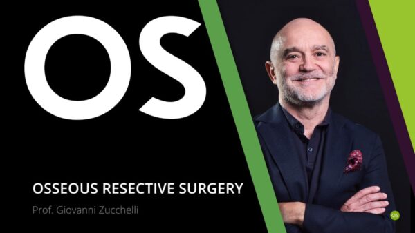
Osteocom Osseous Resective Surgery – Giovanni Zucchelli
Bone Resective Surgery for Esthetic and Functional Clinical Crown Lengthening
In this section, you will find video courses and video lectures focusing on bone resective surgery, a surgical treatment preparatory to your mucogingival and periodontal clinical procedures.
Here you will find everything you need to know about identifying, managing and resolving a wide range of underlying bone defects.
Join our “Resective Surgery” video lectures and make your periodontal surgeries easier, more effective, safer and more predictable.
Professor Giovanni Zucchelli and osteocom welcome you to this new video course
that will delve into the world of osseous resective surgery.
You will receive a comprehensive and specialized training on how to perform an
effective, safe and predictable clinical crown lengthening to enhance your patients’
smile aesthetics and functionality.
Osseous resective surgery procedures shown on this course are preparatory to
performing correct clinical crown lengthening because they restore a correct bone
tissue morphology.
Several clinical videos will be presented, allowing you to appreciate the treatment
protocols adopted by Prof. Zucchelli to eliminate anatomical discrepancies and
reshape periodontal tissues.
Find out how to make your mucogingival surgical procedures more efficient and
easier to perform by preliminarily tackling the underlying bone defects thanks to
the osseous resective surgery techniques you will master at the end of this course.
By attending this course, you will deepen your knowledge of:
• Biological width and early restorative phase (clear)
• Clinical crown lengthening and interdental papilla regeneration
• Passive altered eruption diagnosis
• Gummy smile surgical treatment
• Resective surgery in natural dentition
• Distal wedge procedure and the thinned palatal flap
• Apically positioned flap
• Pre-prosthetic osseous resective surgery
At the end of this course, you will be able to:
• Identify and manage your patients’ bone defects
• Eliminate periodontal pockets
• Reshape bone morphology to facilitate future mucogingival surgery
• Perform clinical crown lengthening to improve smile aesthetics and function
• Adopt innovative protocols such as “In&Out” drilling technique and FibRE-ORS
• Correctly understand the anatomical characteristics of soft and hard tissues
• Preventing failures in periodontology
Who the course is addressed to:
The video course is aimed at all professionals who already perform routine
periodontal and implant therapies.
Specifically, the course is particularly recommended for all those who would like to
learn more about anatomical, diagnostic and prognostic concepts for correct
advanced restorative surgical therapy.
This course includes:
• 11 theoretical video lectures with several clinical videos showed and discussed
in detail
Video Lectures:
Video Lecture 1 – Clinical crown lengthening osteoplasty indications (45min)
Description:
In this first video lecture, Prof. Zucchelli will discuss the two areas of resective
surgery on natural teeth: osseous resective surgery and clinical crown
lengthening.
He will also discuss in detail the reasons why osteoplasty should be preferred over
the more incorrectly used ostectomy to treat periodontal patients.
In particular, this lesson covers the following topics:
• Which factors should be considered before starting resective surgery on a
natural tooth?
• When to proceed with clinical crown lengthening?
• What is the main goal of resective surgery?
• The importance of osteoplasty to avoid the risk of peri-implantitis
• The advantages of a correct gingivectomy
• Root anatomy radiographic evaluations
Video Lecture 2 – Thinned palatal flap with ridge advancement (50min)
Description:
In this presentation, Prof. Zucchelli will discuss several techniques for apical soft
tissue repositioning through paramarginal incisions and the use of a thinned
palatal flap.
Risks, good and bad clinical habits and the importance of a perfect initial incision
design will be analysed thanks to numerous clinical videos.
In particular, this lesson covers the following topics:
• How to correctly design incision lines
• Differences between horizontal palatal incisions and the vertical ones
• How to apically replace vertical palate
• Factors who influence palatal flap design
• The risk of damaging the palatine artery by performing resective surgery
Video Lecture 3 – Osteoplastic treatment for the elimination of the bone wall
defect (15min)
Description:
Thanks to this video lecture participants will learn more about the techniques for a
correct osteoplasty and osteotomy with particular reference to drilling and
suturing protocols.
An innovative drilling technique such as the “In&Out” technique will be discussed.
In particular, this lesson covers the following topics:
• In&Out drilling technique
• Restoration of correct bone architecture through removal of supporting bone
• Osteoplasty phases and creation of bone showers
• Osteotomy to accentuate incision contours and eliminate bone peaks
• Mattress suture technique
Video Lecture 4 – Video Surgery with commentary: osteoplasty treatment for
wall bone defect removal (40min)
Description:
In this lecture a surgical video showing an osteoplasty treatment will be presented.
Prof. Zucchelli’s goal will be the removal of wall bone defect.
Participants will thus see the theoretical knowledge learnt from the previous lessons
put into practice.
In particular, this lesson covers the following topics:
• How to incise the palatine flap
• How to operate a mesial flap
• How to draw and open the surgical papilla area
• How to detach the soft tissue of the secondary palatine flap
• How to separate the distal wedge from the bone plate avoiding vascular
problems
• In&Out drilling technique
Video Lecture 5 – FibRE-ORS surgical technique (10min)
Description:
In this fifth video lecture the FibRE-ORS technique will be introduced.
This innovative methodology involves using the fibres present in the most apical part
of the tooth to determine the In&Out, i.e. the position where the cylindrical flat-head
drill should be inserted to bring the bottom of the infrabony defect to the exterior.
In particular, this lesson covers the following topics:
• Identify the supracrestal connective fibre system underlying the infrabony
defect
• How to reduce post-operative sensitivity by using ultrasonic tips to grind the
tooth
• What to do when the fibres are inserted into the base of the infrabony defect
Video Lecture 6 – Biological width and supracrestal attachment tissue (1h)
Description:
Three weeks after resective surgery, the maximum possible clinical crown
lengthening is achieved.
In the following months the crown lengthening will gradually decrease due to the
formation of the soft tissue rebound.
Prof. Zucchelli therefore explains how convenient it is to take advantage of this
provisional crown lengthening to do quick restorative procedures comfortably and
safely when permanent crown lengthening is not desired
In particular, this lesson covers the following topics:
• Biological width: myth or scientific evidence?
• Why can’t we penetrate the area of the supracrestal attachment tissue?
• How does bone resorption reform the supracrestal attachment?
• What happens when we interfere with the connective attachment zone?
• “The 3mm dogma” in clinical crown lengthening therapy
• Soft tissue rebound in the interproximal area
Video Lecture 7 -Factors influencing papillae growth (30min)
Description:
In this video lecture Prof. Zucchelli will discuss factors influencing papillae growth
between two prosthetic elements.
Different positive and negative possible scenarios will be presented with clinical
videos with detailed commentary.
In particular, this lesson covers the following topics:
• How to cause spontaneous convergence between abutments
• Abutment characteristics influencing convergence
• Creating a convergent space for papillae to grow
• Preparation of abutments and relining temporaries
• Case finalization and after surgery Follow-Up
Video Lecture 8 – Single-tooth clinical crown lengthening (45min)
Description:
Single tooth clinical crown lengthening is a resective surgery that requires great
attention to the adjacent teeth.
In this video lesson, a surgical video with commentary by Prof. Zucchelli for single
tooth crown lengthening with major osteoplastic treatment and minimal
ostectomy will be presented.
In particular, this lesson covers the following topics:
• Incision protocol to remove the secondary palatal flap
• Drilling protocol with different size and grain types
• Reducing buccolingual and papilla bone thickness
• Importance of osteoplastic treatment
• Prevent excessive soft tissue regrowth
Video Lecture 9 – Clinical video: Perio-Restorative Surgery (2h 45min)
Description:
In this extensive video lecture, Prof. Zucchelli will explore in detail 3 different
approaches that characterize perio-restorative surgery by presenting a video
resective surgery in the frontal and lateral area.
A specific focus will be given to the relationship between surgeon and prosthetist.
In particular, this lesson covers the following topics:
• Why should the treatment option with intraoperative preparation of the
abutments be excluded?
• The importance of early preparation with interproximal convergence
modification
Video Lecture 10 – Clinical video: step-by-step resective surgery (50min)
Description:
In this lecture, a case documented by a long surgical video with commentary by
Prof. Zucchelli will be presented.
The treatment will be analysed step by step, from incision techniques to suture
protocols up to prosthetic finalization.
In particular, this lesson covers the following topics:
• The risks of damaging the palatine artery during palatal flap thinning
• Locating and removing the distal wedge
• Intra-sucular incisions to remove the secondary palatal flap
• Removal of vestibular bone in teeth
• The “apical mattress” suture technique to move the flap coronally
Video Lecture 11 – Passive altered eruption: diagnosis and treatment (45min)
Description:
In this last video lecture of the course, Prof. Zucchelli will show an overview of
altered passive eruptions, illustrating their possible causes, diagnostic and
therapeutic processes and different classifications, including a final clinical video as
a teaching example.
In particular, this lesson covers the following topics:
• What is passive altered eruption?
• The possibility of developing gingival recession
• What happens when the maxillary bone grows vertically?
• How to diagnose the passive altered eruption
• Classification of the different types of passive altered eruption
By: Giovanni Zucchelli
11 Videos (37.8 GB) (7H:40M)

