No products in the cart.
- Ophthalmology Books, Retina and Vitreous
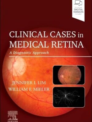
Clinical Cases in Medical Retina: A Diagnostic Approach (Original PDF)
 Ophthalmology Books, Retina and Vitreous
Clinical Cases in Medical Retina: A Diagnostic Approach (Original PDF)
0 out of 5(0)by Jennifer I. Lim MD (Editor), William F. Mieler MD (Editor)
Format : Original PDF
SKU: n/a$10.00 - Ophthalmology Books, Neuro-Ophthalmology
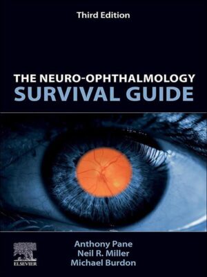
The Neuro-Ophthalmology Survival Guide: The Neuro-Ophthalmology Survival Guide 3rd Edition(EPUB)
 Ophthalmology Books, Neuro-Ophthalmology
The Neuro-Ophthalmology Survival Guide: The Neuro-Ophthalmology Survival Guide 3rd Edition(EPUB)
0 out of 5(0)by Anthony Pane (Author), Neil R. Miller (Author), Michael Burdon (Author)
Format :EPUB
File Size :17 MB
SKU: n/a$15.00 - Dermatology
Rook’s Textbook Of Dermatology, 4 Volume Set, 10th Edition (Original PDF From Publisher)
0 out of 5(0)By Christopher E. M. Griffiths, Jonathan Barker, Tanya O. Bleiker, Walayat Hussain, Rosalind C. Simpson
Language: English
Publisher: Wiley – Blackwell
Edition: 10
Format: Publisher PDF
File Size: 2 GB
SKU: n/a$80.00 - Ophthalmology Books, General Ophthalmology
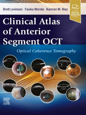
Clinical Atlas Of Anterior Segment OCT: Optical Coherence Tomography (EPub+Converted PDF)
 Ophthalmology Books, General Ophthalmology
Clinical Atlas Of Anterior Segment OCT: Optical Coherence Tomography (EPub+Converted PDF)
0 out of 5(0)by Brett Levinson (Editor), Faskia Woreta (Editor), Kamran Riaz (Editor)
Publisher: Elsevier
Format : EPub+Converted PDF
SKU: n/a$35.00 - Ophthalmology Books, General Ophthalmology
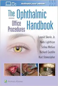
The Ophthalmic Office Procedures Handbook (Epub+Converted PDF)
 Ophthalmology Books, General Ophthalmology
The Ophthalmic Office Procedures Handbook (Epub+Converted PDF)
0 out of 5(0)By Leonid Skorin Jr OD DO, Nathan Robert Lighthizer, Selina McGee, Richard Castillo, Karl Stonecipher
Publisher Lippincott Williams & Wilkins
Edition : 1
Format : EPub+Converted PDF
SKU: n/a$24.00
- Ophthalmology Books, Free Download ophthalmology books
Ophthalmic Medical Image Analysis: 9th International Workshop, OMIA 2022, Held in Conjunction with MICCAI 2022, Singapore, Singapore, September 22, … (Lecture Notes in Computer Science, 13576)
 Ophthalmology Books, Free Download ophthalmology books
Ophthalmic Medical Image Analysis: 9th International Workshop, OMIA 2022, Held in Conjunction with MICCAI 2022, Singapore, Singapore, September 22, … (Lecture Notes in Computer Science, 13576)
0 out of 5(0)by Bhavna Antony (Editor), Huazhu Fu (Editor), Cecilia S. Lee (Editor), Tom MacGillivray (Editor), Yanwu Xu (Editor), Yalin Zheng (Editor)
Format: Original PDF
Size: 40 MB
SKU: n/a - Free Download ophthalmology books, Ophthalmology Books
Eye Yield: Ophthalmology Basics for Board and FRCS Part 1 Exams
- Free Download ophthalmology books, General Ophthalmology
[Free Download] 2018-2019 Basic and Clinical Science Course (BCSC), Complete Set
- Free Download ophthalmology books, General Ophthalmology
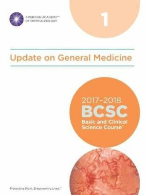
[Free Download] 2017-2018 Basic and Clinical Science Course (BCSC), Complete Print Set
- Free Download ophthalmology books, General Ophthalmology
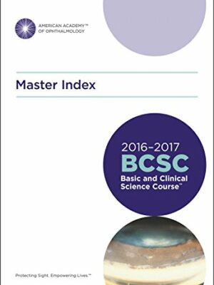
[Free Download] 2016-2017 Basic and Clinical Science Course (BCSC): Complete Set
- Free Download ophthalmology books
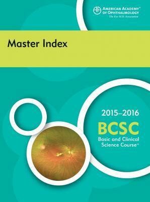
[Free Download] 2015-2016 Basic and Clinical Science Course (BCSC) Complete Set
- Free Download ophthalmology books, General Ophthalmology
A complete CBME guide for Undergraduate and Postgraduate Ophthalmology AWZ3
- Free Download ophthalmology books, General Ophthalmology
Operative Dictations in Ophthalmology, 2nd Edition
- Free Download ophthalmology books, Oculofacial Plastic and Orbital Surgery, Ophthalmic Pathology and Intraocular Tumors, Retina and Vitreous
The Wills Eye Manual: Office and Emergency Room Diagnosis and Treatment of Eye Disease, 6th Edition
- Ophthalmology Books, Retina and Vitreous
Atlas of Retinal OCT: Optical Coherence Tomography 2nd Edition
 Ophthalmology Books, Retina and Vitreous
Atlas of Retinal OCT: Optical Coherence Tomography 2nd Edition
0 out of 5(0)by Jay S. Duker MD (Editor), Nadia K. Waheed MD MPH (Editor), Darin Goldman MD (Editor), Shilpa J. Desai MD (Editor)
Format : EPUB
File Size : 52.9 MB
SKU: n/a$10.00 - Ophthalmology Books, Courses, General Ophthalmology
2023-2024 Basic and Clinical Science Course (BCSC) Complete Set
 Ophthalmology Books, Courses, General Ophthalmology
2023-2024 Basic and Clinical Science Course (BCSC) Complete Set
0 out of 5(0)Publisher: AAO
Format : 13 Original PDFs + Video included (QR code)
File Size : 2.65 GB
SKU: n/a$100.00 - Ophthalmology Books, Cataract & Refractive Surgery, Optometry
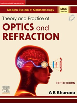
Theory and Practice of Optics & Refraction 5th Edition
 Ophthalmology Books, Cataract & Refractive Surgery, Optometry
Theory and Practice of Optics & Refraction 5th Edition
0 out of 5(0)by A. K. Khurana (Author)
Format: Original PDF
SKU: n/a$15.00 - Ophthalmology Books, Pediatric Ophthalmology and Strabismus
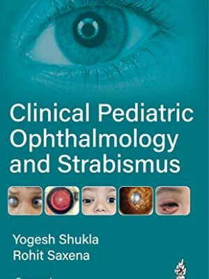
Clinical Pediatric Ophthalmology and Strabismus
 Ophthalmology Books, Pediatric Ophthalmology and Strabismus
Clinical Pediatric Ophthalmology and Strabismus
0 out of 5(0)by Yogesh Shukla (Author), Rohit Saxena (Author)
Format: Original PDF from Publisher
Size: 23.7 MB
SKU: n/a$10.00 - Ophthalmology Books, External Disease and Cornea
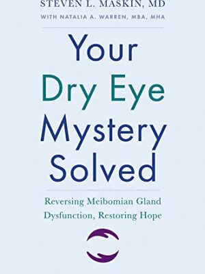
Your Dry Eye Mystery Solved: Reversing Meibomian Gland Dysfunction, Restoring Hope (Yale University Press Health & Wellness)
 Ophthalmology Books, External Disease and Cornea
Your Dry Eye Mystery Solved: Reversing Meibomian Gland Dysfunction, Restoring Hope (Yale University Press Health & Wellness)
0 out of 5(0)by Steven L. Maskin (Author), Natalia A. Warren
Format : EPUB
File Size : 3 MB
SKU: n/a$10.00 - Cataract & Refractive Surgery, Courses, External Disease and Cornea, General Ophthalmology, Glaucoma, Oculofacial Plastic and Orbital Surgery, Ophthalmic Pathology and Intraocular Tumors, Ophthalmology Books, Optometry, Pediatric Ophthalmology and Strabismus, Retina and Vitreous, Uveitis and Ocular Inflammation
Osler Ophthalmology Online Review 2022
 Cataract & Refractive Surgery, Courses, External Disease and Cornea, General Ophthalmology, Glaucoma, Oculofacial Plastic and Orbital Surgery, Ophthalmic Pathology and Intraocular Tumors, Ophthalmology Books, Optometry, Pediatric Ophthalmology and Strabismus, Retina and Vitreous, Uveitis and Ocular Inflammation
Osler Ophthalmology Online Review 2022
0 out of 5(0)Publisher: Osler
Format : 54 Videos + 6 PDFs
File Size : 7.09 GB
SKU: n/a$65.00 - General Ophthalmology, Cataract & Refractive Surgery, Ophthalmic Pathology and Intraocular Tumors
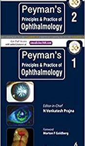
[Original PDF] Peyman’s Principles and Practice of Ophthalmology, 2nd edition, Two Volume Set
 General Ophthalmology, Cataract & Refractive Surgery, Ophthalmic Pathology and Intraocular Tumors
[Original PDF] Peyman’s Principles and Practice of Ophthalmology, 2nd edition, Two Volume Set
0 out of 5(0)Peyman’s Principles and Practice of Ophthalmology, 2nd edition, Two Volume Set
By N Venkatesh Prajna
SKU: n/a$25.00
- Dermatology
Rook’s Textbook Of Dermatology, 4 Volume Set, 10th Edition (Original PDF From Publisher)
0 out of 5(0)By Christopher E. M. Griffiths, Jonathan Barker, Tanya O. Bleiker, Walayat Hussain, Rosalind C. Simpson
Language: English
Publisher: Wiley – Blackwell
Edition: 10
Format: Publisher PDF
File Size: 2 GB
SKU: n/a$80.00 - Courses, General Ophthalmology, Ophthalmology Books, Pathology, USMLE
Osler Ophthalmology 2023 Subscription-Based Review
 Courses, General Ophthalmology, Ophthalmology Books, Pathology, USMLE
Osler Ophthalmology 2023 Subscription-Based Review
0 out of 5(0)By The Osler Institute
6 PDFs + 57 HD Videos
Size: 7 Gb
Download or watch online
SKU: n/a$70.00 - Gastroenterology, Oncology-Hematology, Pathology, Psychiatry, USMLE
PASS-Program USMLE and COMLEX Step 1 – Level 1 CK OnDemand Videos 2023 (VIDEOS)
 Gastroenterology, Oncology-Hematology, Pathology, Psychiatry, USMLE
PASS-Program USMLE and COMLEX Step 1 – Level 1 CK OnDemand Videos 2023 (VIDEOS)
0 out of 5(0)SKU: n/a$55.00 - Dermatology, Gastroenterology, Oncology-Hematology, Orthopedics, Rheumatology, USMLE
Uworld STEP 3 QBank Updated August 2023 (PDF) Systemwise
 Dermatology, Gastroenterology, Oncology-Hematology, Orthopedics, Rheumatology, USMLE
Uworld STEP 3 QBank Updated August 2023 (PDF) Systemwise
0 out of 5(0)SKU: n/a$40.00 - Dermatology, Gastroenterology, Neurology, Rheumatology, Urology, USMLE
TrueLearn USMLE Step 1 Question Bank (Updated Feb 2023) (PDF) Specialty-wise
 Dermatology, Gastroenterology, Neurology, Rheumatology, Urology, USMLE
TrueLearn USMLE Step 1 Question Bank (Updated Feb 2023) (PDF) Specialty-wise
0 out of 5(0)SKU: n/a$10.00

