No products in the cart.
Radiology
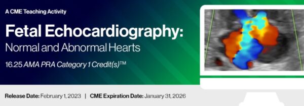
2023 Fetal Echocardiography: Normal and Abnormal Hearts – A Video CME Teaching Activity
CME Release Date 3/01/2022
$35.00
This CME teaching activity provides detailed information on the evaluation of the normal fetal heart both from an anatomic and functional approach. Detailed information on the use of three-dimensional ultrasound in fetal echocardiography and cardiac imaging in the early gestation is presented. This activity also provides comprehensive evaluation of various cardiac malformations, involving abnormalities of the cardiac chambers and the outflow tracts.
Release Date : 1 Feb 2023
Target Audience
This course will greatly benefit physicians and sonographers in the fields of obstetric imaging, fetal echocardiography and pediatric cardiology. Residents in obstetrics and gynecology, as well as fellows in maternal fetal medicine and pediatric cardiology, will also benefit greatly. It should also prove valuable to those physicians and sonographers wanting to gain a more in-depth knowledge of fetal echocardiography
Educational Objectives ▼
At the completion of this CME teaching activity, you should be able to:
Discuss indications for fetal echocardiography, review existing guidelines and the approach to patient counseling.
Optimize ultrasound equipment for the use of 2D and Doppler in fetal echocardiography.
Review the normal fetal cardiac anatomy.
Describe the abnormalities of the venous system of the fetal heart.
Discuss in detail anomalies of the cardiac chambers to include septal defects and anomalies of the right and left ventricle.
Review in detail anomalies of the outflow tracts to include transposition of great arteries, tetralogy of Fallot complex and aortic arch abnormalities.
No special educational preparation is required for this CME activity.
37 Videos
Topics
Current Indications for Fetal Echocardiography: The New National Guidelines
How to Optimize Your Image in Fetal Cardiac Ultrasound
Normal Anatomy of the Cardiac Chambers
Cardiac Chambers Abnormalities: Anomalies of the Right Ventricle
Case Report 1
Cardiac Chambers Abnormalities: Atrioventricular Septal Defect
Cardiac Chambers Abnormalities: The Fetal Borderline Left Ventricle
Cardiac Chambers Abnormalities: Hypoplastic Left Heart Syndrome
Cardiac Chambers Abnormalities: Single Ventricle Anatomy – Type Congenital Heart Disease
Hands-on Scanning Session 1: Optimizing Your Fetal Echo Examination
Congenital Heart Disease in Multiple Pregnancies
Cardiac Tumors
The Placenta and Congenital Heart Disease
Case Report 2
Panel Discussion – Question and Answer Session: Normal Cardiac Anatomy and Cardiac Chamber Abnormalities
The Developing Fetal Brain in Congenital Heart Disease
Screening for Cardiac Malformations in the First and Second Trimester of Pregnancy
The Three-Vessel Trachea View
Anomalies of the Great Vessels: Transposition of the Great Arteries
Case Report 3
Anomalies of the Great Vessels: Tetralogy of Fallot
Anomalies of the Great Vessels: Aortic Arch Abnormalities
Ultrasound Evaluation of Situs Abnormalities
Oxygen for Fetal Intervention in Congenital Heart Disease: What is the Evidence?
Approach to Diagnosis of Systemic Venous Malformations
Approach to Diagnosis of Pulmonary Venous Malformations
Approach to Diagnosis and Management of Fetal Rhythm Abnormalities
Counseling for Prenatal Congenital Heart Disease
Case Report 4
The Genetics of Cardiac Malformations
Long QT Syndrome: Prenatal Diagnosis and Management
Cardiovascular Malformations of Twin-Twin Transfusion Syndrome
Fetal Cardiac Imaging in the First Trimester
Case Report 5
Hands-on Scanning 2: Review of Normal Cardiac Anatomy
Review of Fetal Cardiac Malformations: Putting it All Together
Case Report 6
Samples
https://www.docmeded.com/video/item/8639


