No products in the cart.
+ Include: 26 videos + 1 pdf, size: 6.67 GB
+ Target Audience: radiologists, neurologists and neurosurgeons
Released : 08/15/23
About This CME Teaching Activity ▼
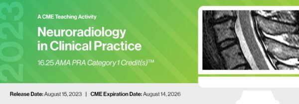
Neuroradiology in Clinical Practice addresses current topics in neuroimaging and the clinical management of neurological disease. Emphasis is placed on the role of imaging in pathology and trauma affecting the central nervous system. Topics in this activity include imaging manifestation of disease, how to optimize imaging techniques and review of the recent advanced applications of artificial intelligence (AI), traumatic brain injury and stroke imaging updates.
Target Audience ▼
This program is designed for practicing radiologists, neurologists and neurosurgeons that interpret or rely on imaging studies for the evaluation of neurological disorders.
Scientific Sponsor ▼
Educational Symposia
Educational Objectives ▼
At the completion of this CME teaching activity, you should be able to:
Discuss the advances in neuroimaging applications and interpretation.
Evaluate intracranial tumors, vascular abnormalities and traumatic brain injury.
Discuss the clinical and imaging manifestations of neurodegenerative disease.
Recognize image characteristics of common neurovascular disease processes.
Optimize CTA and MRA imaging protocols in neuroradiology.
Describe the changing role of imaging in the management of spine disease.
Explain the clinical impact of artificial intelligence in neuroradiology.
+ Topics:
AI and Image Reconstruction Lawrence N. Tanenbaum, M.D., FACR
Multimodal Imaging of Non-Aneurysmal, Nontraumatic Intracranial Hemorrhage J. Pablo Villablanca, M.D., FACR
Optimizing Imaging of the Instrumented Spine Lawrence N. Tanenbaum, M.D., FACR
Imaging Cranial Neuropathy: I-VI Blake A. Johnson, M.D., FACR
Updates in Stroke Imaging Max Wintermark, M.D., MAS, MBA
Dementia Imaging: What to Remember Suzie Bash, M.D.
Imaging Cranial Neuropathy: VII – XII Blake A. Johnson, M.D., FACR
Imaging of Carotid Artery Disease Max Wintermark, M.D., MAS, MBA
Quantitative Volumetric Imaging: Size Matters Suzie Bash, M.D.
Evaluation of Pulsatile Tinnitus William J. Garvis, M.D.
AI in Spine Imaging Lawrence N. Tanenbaum, M.D., FACR
Perfusion Imaging Max Wintermark, M.D., MAS, MBA
Imaging CNS Infections J. Pablo Villablanca, M.D., FACR
Imaging of Traumatic Brain Injury Max Wintermark, M.D., MAS, MBA
PET/MRI in Neuroimaging: Essential or Extravagant? Suzie Bash, M.D.
Facial Canal-lateral Canal: Third Window Pathology William J. Garvis, M.D.
Differential Diagnosis of Basal Ganglia Abnormalities Blake A. Johnson, M.D., FACR
Headache: When to Image and What to Look For Jeffrey S. Ross, M.D.
CT Temporal Bone Features Important to Otology/Neurology William J. Garvis, M.D.
Technology and Empowerment in the Imaging Enterprise Lawrence N. Tanenbaum, M.D., FACR
Imaging of Spine Tumors: Intradural Jeffrey S. Ross, M.D.
COVID and the CNS: What the Radiologist Needs to Know Blake A. Johnson, M.D., FACR
Spontaneous Intracranial Hypotension: Diagnosis and Treatment of Spinal Leaks Jeffrey S. Ross, M.D.
Neuroimaging of Pain: Visualizing the Invisible J. Pablo Villablanca, M.D., FACR
Spine Infection and Inflammatory Disease Jeffrey S. Ross, M.D.
Spine Imaging: Pearls and Pitfalls – Syrinx?, Transitional?, Unstable? and More J. Pablo Villablanca, M.D., FACR


