No products in the cart.
Dentistry, Internal Medicine, Pathology, Radiology
Oakstone Society for Vascular Medicine Comprehensive Review of Vascular Ultrasound Interpretation and Registry Preparation 2023
Format : 28 Videos + 2 PDFs
$35.00
Description
+ Include: videos + pdf, size: GB
+ Target Audience: cardiologists, vascular medicine specialists, vascular surgeons, vascular technologists, vascular and general sonographers, radiologists
+ Information:
This online CME program spans 40 lectures that expose you to all aspects of non-invasive vascular testing. Abundant case study materials and over 100 registry-type exam questions will help those who work within the non-invasive vascular laboratory to prepare for the RPVI examination and maintain IAC Vascular Testing continuing medical education requirements. Nationally-renowned speakers provide expanded content on vascular lab operations and ergonomics, as well as valuable insight on:
Imaging techniques (grayscale, color and spectral Doppler, physiological testing)
Various testing protocols and diagnostic criteria
A broad range of vascular pathology, both common and rare
Fundamentals of vascular physics
Quality improvement and patient safety
And more…
Date of Original Release: June 30, 2023
Date Credits Expire: June 29, 2026
CME credit is obtained upon successful completion of an online activity post-test and evaluation.
Unique Learning Objectives
At the completion of this course, you should be able to:
Recognize basic ultrasound physics concepts and their application to vascular ultrasound and physiologica testing
Identify common imaging and Doppler artifacts encountered in the vascular laboratory
Use grayscale imaging, color Doppler analysis, and spectral Doppler waveforms to assist in the diagnosis of arterial and venous disease
Apply interpretation skills for diagnosis of internal carotid artery stenosis using duplex ultrasound
Apply interpretation skills for diagnosis of venous thrombosis and venous valvular reflux using duplex ultrasound
Use arterial duplex and physiological testing to assess severity and anatomic location of lower extremity arterial disease
Apply standard diagnostic criteria to diagnose abdominal aortic aneurysm (AAA) and renal and mesenteric artery stenosis using duplex ultrasound
Use color and spectral Doppler analysis to assess the patency of arterial and venous stents and following endovascular AAA repair
Recognize uncommon and rare vascular disorders encountered in the vascular laboratory
Use best practices for running a high quality vascular laboratory, including quality improvement program, accreditation, and prevention of work-related musculoskeletal disorder (WRMSD) among technical personnel
Identify areas of knowledge deficit to improve preparation for the ARDMS Registered Physician in Vascular Interpretation (RPVI) examination
Intended Audience
This program is designed for cardiologists, vascular medicine specialists, vascular surgeons, vascular technologists, vascular and general sonographers, radiologists, nurse practitioners, and physician assistants.
+ Topics:
Basics of Laboratory Technology and Operations
Preparing for the Registry Examination – Heather L. Gornik, MD, RVT, RPVI, MSVM
Physics and Instrumentation I – Fredrick Kremkau, PhD
Physics and Instrumentation II – Fredrick Kremkau, PhD
Transducer Selection, Image Optimization, Spectral and Color Doppler, and B-Mode Artifacts – Ann Marie Kupinski, PhD, RVT, RDMS, FSVU
Quality Improvement in the Vascular Laboratory – Heather L. Gornik, MD, RVT, RPVI, MSVM
Fundamentals of Ergonomics in the Vascular Lab – Jill Sommerset, RVT
Cerebrovascular
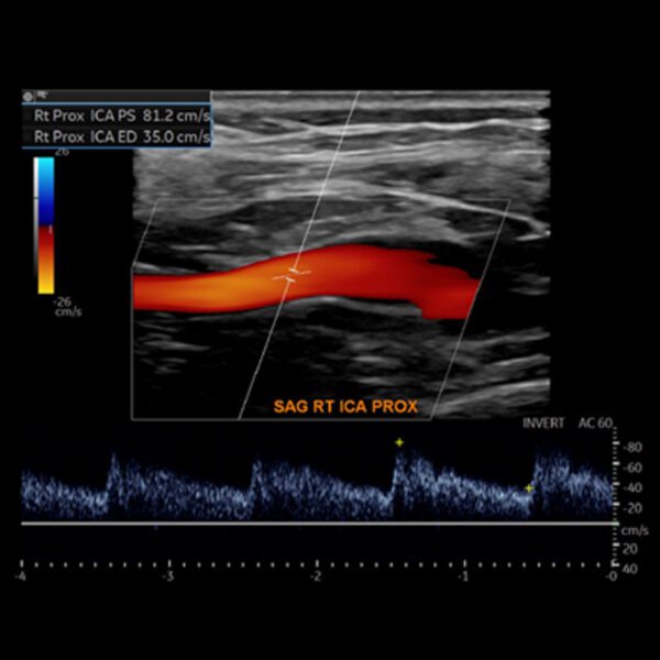
Basics of the Carotid Duplex Examination and Criteria for Diagnosis of Internal Carotid Artery Stenosis – Heather L. Gornik, MD, RVT, RPVI, MSVM
Carotid Evaluation Following Stents and Endarterectomy, Interpretive Pitfalls – R. Eugene Zierler, MD, RPVI, FACS, FSVM
Non-atherosclerotic Cerebrovascular Disease – Duplex Findings – Daniella Kadian-Dodov, MD, RPVI, FSVM
Aortic Arch Vessel and Vertebral Artery Findings, Subclavian Steal – Daniella Kadian-Dodov, MD, RPVI, FSVM
Transcranial Doppler Essentials – Larry Raber, RVT, RDMS, RT
Interpretive Case Review – Carotid, Vertebral, and Subclavian Arteries – Rapid Fire Cases – Aditya Sharma, MBBS, RPVI, FSVM
Peripheral Arterial
Lower Extremity Arterial Physiological Testing – Ana Casanegra, MD, RPVI, FSVM
Lower Extremity Arterial Duplex – R. Eugene Zierler, MD, RPVI, FACS, FSVM
Upper Extremity Arterial Testing – Marie D. Gerhard Herman, MD, RVT, RPVI
Pedal Artery Duplex and CLTI Duplex Imaging – Jill Sommerset, RVT
Arterial Access Complications – Natalia Fendrikova-Mahlay, MD, RPVI
Dialysis Access Mapping and Post Procedure Evaluation – Ann Marie Kupinski, PhD, RVT, RDMS, FSVU
Interpretive Case Review – Physiologic Testing and Duplex of Native Upper and Lower Extremity Arteries – Rapid Fire Cases – Aditya Sharma, MBBS, RPVI, FSVM
Interpretive Case Review – Duplex Assessment of Arterial Bypass Grafts and Stents – Rapid Fire Cases – Ido Weinberg, RPVI, FSVM
Interpretive Case Review – Access Complications, Dialysis Access, Vascular Zebras – Rapid Fire Cases – Jeffrey Olin, DO, RPVI, MSVM
Abdominal
Renal (Native and Transplant) Duplex Ultrasound, Renal Stents – Natalie Evans, MD, RPVI, FSVM
Mesenteric Artery Duplex Ultrasound, Mesenteric Stents – Natalia Fendrikova-Mahlay, MD, RPVI
Assessment of the Aorta, Surveillance of AAA, and Follow-up of Aortic Endografts – R. Eugene Zierler, MD, RPVI, FACS, FSVM
Duplex Doppler Assessment of Hepatic-Portal Vasculature – Nirvikar (Nirvi) Dahiya, MD
Interpretive Case Review – Abdominal Imaging – Abdominal Aorta, AAA; and Endografts; Renal and Mesenteric Ultrasound – Rapid Fire Cases – Natalie Evans, MD, RPVI, FSVM
Interpretive Case Review – Abdominal Imaging – Aortic, Mesenteric, Renal and Pelvic Arterial and Venous Compression Syndromes, Arteriopathies – Rapid Fire Cases – Jeffrey Olin, DO, RPVI, MSVM
Peripheral Venous
Venous Duplex for Diagnosis of Deep Venous Thrombosis – Marie D. Gerhard Herman, MD, RVT, RPVI
Bare Bones for the Boards – Venous Physiological Testing – Ana Casanegra, MD, RPVI, FSVM
Venous Duplex for Assessment of Venous Valvular Incompetency, Mapping for Endovenous Therapies, Assessing for EHIT – Raghu Kolluri, MD, RVT, FSVM
Venous Duplex for Assessment of Central Veins – Karem Harth, MD
Interpretive Case Review – Venous Testing 1 – Rapid Fire Cases – Karem Harth, MD
Interpretive Case Review – Venous Testing 2 – Rapid Fire Cases – Raghu Kolluri, MD, RVT, FSVM
Mock RPVI Examinations and Additional Talks
Key Non-Vascular Incidental Findings Every Reader Should Know – Nirvikar (Nirvi) Dahiya, MD
Physics, Technology, and Instrumentation Mock RPVI Examination – Ann Marie Kupinski, PhD, RVT, RDMS, FSVU
Mock Examination Questions – Session I – Randy Ramcharitar, MD
Mock Examination Questions – Session II – Ido Weinberg, RPVI, FSVM
Mock Examination Questions – Session III – Aditya Sharma, MBBS, RPVI, FSVM
Mock RPVI Examination Questions – TCD and TCI – Larry Raber, RVT, RDMS, RT
Mock Examination Questions – Essentials of Accreditation, Patient Care and Safety, Quality Assurance, and Test Validation – Heather L. Gornik, MD, RVT, RPVI, MSVM


