No products in the cart.
Description
CME: Abdominal and Pelvic Imaging
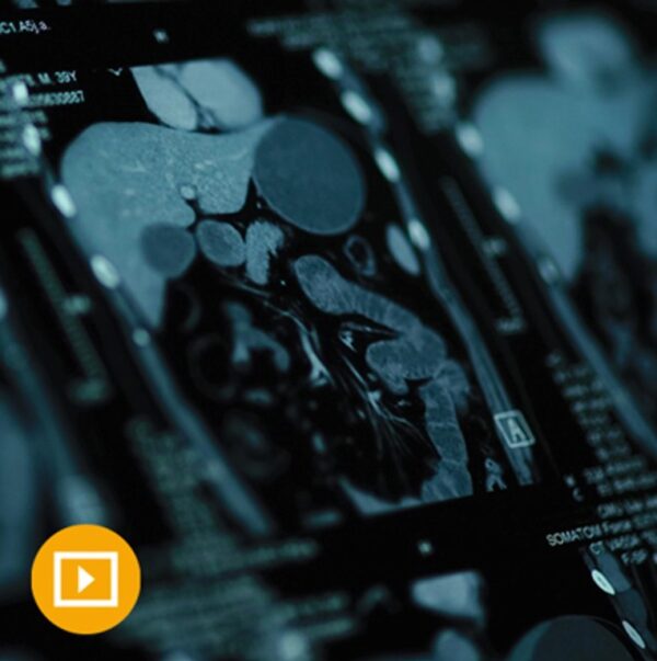
Through this online video CME program, you’ll explore state-of-the-art methods and evidence-based practices for gastrointestinal and genitourinary imaging studies. Expert faculty also review ACR guidelines for managing incidental findings on CT scans of the kidney, adrenal, liver and pancreas.
UCSF Abdominal and Pelvic Imaging is convenient continuing medical education that will help you to better:
Identify indications for the use of different imaging modalities (CT/MRI/US) for various abdomino-pelvic organs and conditions
Apply practical approaches to diagnosing both benign and malignant diseases of the abdomen and pelvis
Anticipate potential pitfalls in abdomino-pelvic imaging and interpretation
And more!
Accreditation
The University of California, San Francisco School of Medicine (UCSF) is accredited by the Accreditation Council for Continuing Medical Education (ACCME) to provide continuing medical education for physicians.
Designation
UCSF designates this educational activity for a maximum of 14.00 AMA PRA Category 1 Credits™. Physicians should claim only the credit commensurate with the extent of their participation in the activity.
The total credits are inclusive of 5.00 in CT, 5.00 in MR, 4.00 in US, and 1.00. in PET.
Series Release: May 1, 2023
Series Expiration: April 30, 2026 (deadline to register for credit)
Estimated Time to Complete Activity: 14.00 hours
CME credit is obtained upon successful completion of an activity post-test and evaluation. CME Credit registration forms must be submitted prior to series expiration date. Certificates will be dated upon receipt and cannot be dated retroactively.
Learning Objectives
At the conclusion of this activity, the participant will be able to:
Prioritize management of incidental findings
Identify indications for the use of different imaging modalities (CT/MRI/US) for various abdomino-pelvic organs and conditions
Apply practical approaches to diagnosing both benign and malignant diseases of the abdomen and pelvis
Anticipate potential pitfalls in abdomino-pelvic imaging and interpretation
+ Topics:
CT/MRI Focal Hepatic Lesions – Part 1 – Spencer Behr, MD
Pancreatic Cancer Imaging – Pearls and Pitfalls – Jane Wang, MD
Beyond the Solid Organs – Mesentery, Omentum & Peritoneum – Jane Wang, MD
Incidental Pancreatic Cystic Lesions – Review of Management Guidelines and Controversies – Jane Wang, MD
FDG PET – Pearls and Pitfalls of Recess – Spencer Behr, MD
Rectal Cancer Staging – Jane Wang, MD
Incidental Renal Masses – What to Do Next – Jane Wang, MD
Small Bowel Emergencies – Jane Wang, MD
Interesting GI/GU Cases – Jane Wang, MD
Retroperitoneal Masses – Spencer Behr, MD
Abdominal Organ Transplant 1 – Living Donor Evaluation and Transplant Configurations – Joseph Leach, MD, PhD
CT – MRI Focal Hepatic Lesions – Part 2 – HCC and LI‐RADS – Spencer Behr, MD
Acute Pelvic Pain – Negative Pregnancy Test – Liina Poder, MD
Abdominal Organ Transplant 2 – Vascular Complications – Joseph Leach, MD, PhD
Acute Pelvic Pain: Positive Pregnancy Test – Liina Poder, MD
Abdominal Vascular Disease and Acute Injury – Joseph Leach, MD, PhD
Prostate Specific Membrane Antigen – PET – Spencer Behr, MD
Vascular MRI – Techniques and Tips – Joseph Leach, MD PhD
How to Work‐up Malignancy in Pregnancy – Liina Poder, MD
Arterial Ultrasound – Cervical and Extremities – Joseph Leach, MD PhD
Just Say Yes – An Inspirational Guide to Procedures for the General Radiologist – Liina Poder, MD
Adrenal Imaging – Spencer Behr, MD
MRI for Endometriosis – Liina Poder, MD
Adnexa – Multimodality Approach – Liina Poder, MD
Vascular Imaging – Multimodality Case‐based Review – Joseph Leach, MD, PhD
Retained Products of Conception – How to Approach the Conundrum – Liina Poder, MD
Interesting Abdominal Cases Through the Years – Spencer Behr, MD


