No products in the cart.
- Ophthalmology Books, Glaucoma
Research Protocols for Ophthalmic Disease Mechanisms and Therapeutics: Glaucoma – Ocular Hypertension – Original PDF
 Ophthalmology Books, Glaucoma
Research Protocols for Ophthalmic Disease Mechanisms and Therapeutics: Glaucoma – Ocular Hypertension – Original PDF
0 out of 5(0)Research Protocols for Ophthalmic Disease Mechanisms and Therapeutics: Glaucoma – Ocular Hypertension
Sharif, Najam A.Ohia, Sunny E.
Bentham Science Publishers 2025Original PDF
SKU: n/a - Ophthalmology Books, General Ophthalmology
Ophthalmic Drug Delivery: Advancement, Industrialization Prospect and Applications
 Ophthalmology Books, General Ophthalmology
Ophthalmic Drug Delivery: Advancement, Industrialization Prospect and Applications
0 out of 5(0)by Neelesh Kumar Mehra PhD (Editor)
Download Format: PDF, EPUB
SKU: n/a - Ophthalmology Books, General Ophthalmology
Kanski. Oftalmología clínica: Un enfoque sistemático, 10th Edition (True PDF from Publisher)
 Ophthalmology Books, General Ophthalmology
Kanski. Oftalmología clínica: Un enfoque sistemático, 10th Edition (True PDF from Publisher)
0 out of 5(0)By John F. Salmon MD FRCS FRCOphth
Format : True PDF, Cover, Bookmark, ToC, Index
File Size : 133.8SKU: n/a - Ophthalmology Books, Retina and Vitreous
Rétinopathie diabétique, 3rd Edition (EPUB)
0 out of 5(0)by Pascale Massin, Ali Erginay, Bénédicte Dupas, Sylvie Feldman, Amélie Lecleire-Collet, Michel Paques, Sophie Bonnin, Catherine Vignal-Clermont, Aude Couturier, Mohamed Bennani
Format : EPUB
File Size : 54.2SKU: n/a - Ophthalmology Books, Retina and Vitreous
Rétine : atlas des maladies du fond d’oeil (French Edition) (True PDF from Publisher)
 Ophthalmology Books, Retina and Vitreous
Rétine : atlas des maladies du fond d’oeil (French Edition) (True PDF from Publisher)
0 out of 5(0)By Salomon Yves Cohen, Prithvi RAMTOHUL, Alain Gaudric, Alexander J. Brucker
Format : True PDF, Cover, Bookmark, ToC, Index
File Size : 76.9SKU: n/a - Ophthalmology Books, General Ophthalmology
Eye Melanoma Unveiled: Cutting-Edge Diagnosis, Revolutionary Treatments and Advanced Drug Delivery (True PDF from Publisher)
 Ophthalmology Books, General Ophthalmology
Eye Melanoma Unveiled: Cutting-Edge Diagnosis, Revolutionary Treatments and Advanced Drug Delivery (True PDF from Publisher)
0 out of 5(0)By Bhupendra G. Prajapati PhD, Devesh U. Kapoor PhD., Nemat Ali PhD.
Format : True PDF, Cover, Bookmark, ToC, Index
File Size : 13.3SKU: n/a - Ophthalmology Books, General Ophthalmology, Oculofacial Plastic and Orbital Surgery
Clinical Ophthalmic Oncology: Eyelid Tumors – Original PDF
 Ophthalmology Books, General Ophthalmology, Oculofacial Plastic and Orbital Surgery
Clinical Ophthalmic Oncology: Eyelid Tumors – Original PDF
0 out of 5(0)by Bhupendra C.K. Patel (Editor), Jacob Pe’er (Editor), Arun D. Singh (Editor)
SKU: n/a - Ophthalmology Books, Retina and Vitreous
Clinical Ophthalmic Oncology: Retinal Tumors Fourth Edition – Original PDF
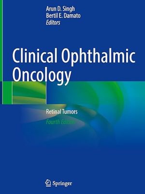 Ophthalmology Books, Retina and Vitreous
Clinical Ophthalmic Oncology: Retinal Tumors Fourth Edition – Original PDF
0 out of 5(0)by Arun D. Singh (Editor), Bertil E. Damato (Editor)
SKU: n/a - Ophthalmology Books, General Ophthalmology, Ophthalmic Pathology and Intraocular Tumors
Clinical Ophthalmic Oncology: Conjunctival Tumors 4th Edition – Original PDF
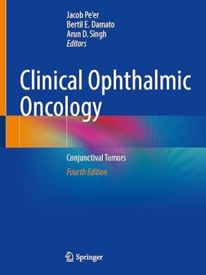 Ophthalmology Books, General Ophthalmology, Ophthalmic Pathology and Intraocular Tumors
Clinical Ophthalmic Oncology: Conjunctival Tumors 4th Edition – Original PDF
0 out of 5(0)by Jacob Pe’er (Editor), Bertil E. Damato (Editor), Arun D. Singh (Editor)
SKU: n/a
- Free Download ophthalmology books, General Ophthalmology
[Free Download] 2018-2019 Basic and Clinical Science Course (BCSC), Complete Set
![Ophthalmology Books, Optometry Books - PDF EPUB 14 [Free Download] 2018-2019 Basic and Clinical Science Course (BCSC), Complete Set](https://ophthalmologyebooks.store/wp-content/uploads/2022/12/2018-2019-Basic-and-Clinical-Science-Course-complete-set-300x400.jpeg)
- Free Download ophthalmology books, General Ophthalmology
[Free Download] 2017-2018 Basic and Clinical Science Course (BCSC), Complete Print Set
![Ophthalmology Books, Optometry Books - PDF EPUB 15 [Free Download] 2017-2018 Basic and Clinical Science Course (BCSC), Complete Print Set](https://ophthalmologyebooks.store/wp-content/uploads/2022/12/3242-300x400.jpeg)
- Free Download ophthalmology books, General Ophthalmology
[Free Download] 2016-2017 Basic and Clinical Science Course (BCSC): Complete Set
![Ophthalmology Books, Optometry Books - PDF EPUB 16 [Free Download] 2016-2017 Basic and Clinical Science Course (BCSC): Complete Set](https://ophthalmologyebooks.store/wp-content/uploads/2022/12/Free-Download-2016-2017-Basic-and-Clinical-Science-Course-BCSC-Complete-Set--300x400.jpeg)
- Free Download ophthalmology books
[Free Download] 2015-2016 Basic and Clinical Science Course (BCSC) Complete Set
![Ophthalmology Books, Optometry Books - PDF EPUB 17 [Free Download] 2015-2016 Basic and Clinical Science Course (BCSC) Complete Set](https://ophthalmologyebooks.store/wp-content/uploads/2022/12/2015-2016-Basic-and-Clinical-Science-Course-298x400.jpeg)
- Free Download ophthalmology books, General Ophthalmology
A complete CBME guide for Undergraduate and Postgraduate Ophthalmology AWZ3
- Free Download ophthalmology books, Oculofacial Plastic and Orbital Surgery, Ophthalmic Pathology and Intraocular Tumors, Retina and Vitreous
The Wills Eye Manual: Office and Emergency Room Diagnosis and Treatment of Eye Disease, 6th Edition
- Free Download ophthalmology books, Ophthalmology Books
The Little Eye Book: A Pupil’s Guide to Understanding Ophthalmology Second Edition

- Ophthalmology Books, Ophthalmic Pathology and Intraocular Tumors
Adler’s Physiology of the Eye 12th Edition (Original PDF from Publisher)
 Ophthalmology Books, Ophthalmic Pathology and Intraocular Tumors
Adler’s Physiology of the Eye 12th Edition (Original PDF from Publisher)
0 out of 5(0)By Leonard A Levin MD PhD FARVO, Paul L. Kaufman MD FARVO, Mary Elizabeth Hartnett MD FARVO
Format: Publisher PDF
SKU: n/a - Ophthalmology Books, General Ophthalmology
Kanski’s Clinical Ophthalmology: A Systematic Approach 10th Edition (Original PDF)
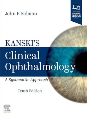 Ophthalmology Books, General Ophthalmology
Kanski’s Clinical Ophthalmology: A Systematic Approach 10th Edition (Original PDF)
0 out of 5(0)By John F. Salmon MD FRCS FRCOphth
Publisher Elsevier
Edition : 10
Format : Original PDF
Size: 67 MB
SKU: n/a - Ophthalmology Books, Optometry
Last-Minute Optics: A Concise Review of Optics, Refraction, and Contact Lenses 3rd Edition (EPub+Converted PDF)
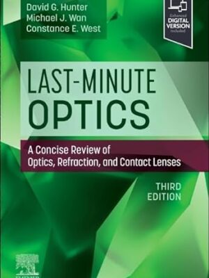 Ophthalmology Books, Optometry
Last-Minute Optics: A Concise Review of Optics, Refraction, and Contact Lenses 3rd Edition (EPub+Converted PDF)
0 out of 5(0)by David G Hunter MD PhD (Author), Michael J. Wan (Author), Constance West (Author)
Publisher: Elsevier
Edition : 3
Format : EPub+Converted PDF
SKU: n/a - Ophthalmology Books, Retina and Vitreous
Atlas of Retinal OCT: Optical Coherence Tomography 2nd Edition
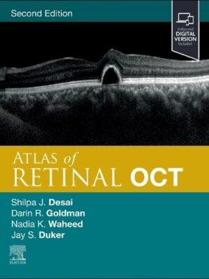 Ophthalmology Books, Retina and Vitreous
Atlas of Retinal OCT: Optical Coherence Tomography 2nd Edition
0 out of 5(0)by Jay S. Duker MD (Editor), Nadia K. Waheed MD MPH (Editor), Darin Goldman MD (Editor), Shilpa J. Desai MD (Editor)
Format : EPUB
File Size : 52.9 MB
SKU: n/a - Ophthalmology Books, Courses, General Ophthalmology
2023-2024 Basic and Clinical Science Course (BCSC) Complete Set
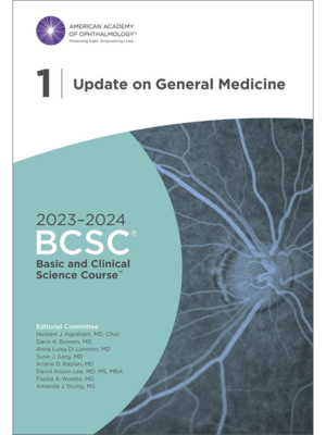 Ophthalmology Books, Courses, General Ophthalmology
2023-2024 Basic and Clinical Science Course (BCSC) Complete Set
0 out of 5(0)Publisher: AAO
Format : 13 Original PDFs + Video included (QR code)
File Size : 2.65 GB
SKU: n/a
- Dermatology
Rook’s Textbook Of Dermatology, 4 Volume Set, 10th Edition (Original PDF From Publisher)
0 out of 5(0)By Christopher E. M. Griffiths, Jonathan Barker, Tanya O. Bleiker, Walayat Hussain, Rosalind C. Simpson
Language: English
Publisher: Wiley – Blackwell
Edition: 10
Format: Publisher PDF
File Size: 2 GB
SKU: n/a - Courses, General Ophthalmology, Ophthalmology Books, Pathology, USMLE
Osler Ophthalmology 2023 Subscription-Based Review
 Courses, General Ophthalmology, Ophthalmology Books, Pathology, USMLE
Osler Ophthalmology 2023 Subscription-Based Review
0 out of 5(0)By The Osler Institute
6 PDFs + 57 HD Videos
Size: 7 Gb
Download or watch online
SKU: n/a - Gastroenterology, Oncology-Hematology, Pathology, Psychiatry, USMLE
PASS-Program USMLE and COMLEX Step 1 – Level 1 CK OnDemand Videos 2023 (VIDEOS)
 Gastroenterology, Oncology-Hematology, Pathology, Psychiatry, USMLE
PASS-Program USMLE and COMLEX Step 1 – Level 1 CK OnDemand Videos 2023 (VIDEOS)
0 out of 5(0)SKU: n/a - Dermatology, Gastroenterology, Oncology-Hematology, Orthopedics, Rheumatology, USMLE
Uworld STEP 3 QBank Updated August 2023 (PDF) Systemwise
 Dermatology, Gastroenterology, Oncology-Hematology, Orthopedics, Rheumatology, USMLE
Uworld STEP 3 QBank Updated August 2023 (PDF) Systemwise
0 out of 5(0)SKU: n/a

